Anatomy Of Chest And Ribs / Introduction Anatomy Thoracic The Gap Physio - Spiral ct of thoracic inlet.. This type of ct scan uses a lower radiation level than a conventional. Each rib wraps around the lung and descends approximately 3 to 5 inches. The purpose of this study was to explore the effect of. How these parts interrelate through joints is described also. Related posts of chest bone anatomy.
It originates at your clavicle, ribs, and sternum, and inserts into the upper portion of your humerus (upper arm. Each rib wraps around the lung and descends approximately 3 to 5 inches. Paschalides medical publications, 2004, with. The chest can be split into two parts; Posteriorly, the heads of the ribs interdigitate with the vertebrae and are numbered according to the inferior vertebra.
The Anatomy Of The Ribs And The Sternum And Their Relationship To Chest Wall Structure And Function Sciencedirect from ars.els-cdn.com The embryologic and anatomic basis of modern surgery. ■ identify the basic anatomy seen on a chest radiograph. Manubrium anteriorly, rib 1 laterally, thoracic vertebrae post… xiphoid process anteriorly, costal cartilages 7 to 10 and rib… What are the features of ribs? The first seven are connected behind with the vertebral column. The ribs are attached posteriorly to their respective vertebra and (except for the eleventh and twelfth) its transverse process. Insert contains images of a typical rib and the first rib. They also have a role in ventilation;
They are twelve in number on either side;
The anatomical structure of the 24 ribs in the human body is complex because of the irregular shape and different lengths of each rib. Continue scrolling to read more below. Learn about chest anatomy with free interactive flashcards. Human anatomy for muscle, reproductive, and skeleton. The purpose of this study was to explore the effect of. Pathology of the heart, mediastinum, lungs and pleura. Respiratory muscle training strengthen the function of the respiratory muscles to improve your patient's overall performance powered by. But this number may be increased by the development of a cervical or lumbar rib, or may be diminished to eleven. Posteriorly, the heads of the ribs interdigitate with the vertebrae and are numbered according to the inferior vertebra. Related online courses on physioplus. The pectoralis major and minor. This type of ct scan uses a lower radiation level than a conventional. It describes the theatre of events.
One that claims that you can't focus on specific parts of your chest (eg. Chest blunt trauma (cbt) and the resultant rib fractures often lead to thoracic collapse. Bone on hand and foot diagram quiz. It discusses the specific anatomy of the ribs and costal cartilages, along with the sternum. It is enclosed by the ribs, the vertebral column, and the sternum, or breastbone, and is separated from the abdominal cavity by the diaphragm.
Chest Bone Anatomy Anatomy Drawing Diagram from image.slidesharecdn.com It discusses the specific anatomy of the ribs and costal cartilages, along with the sternum. Posteriorly, the heads of the ribs interdigitate with the vertebrae and are numbered according to the inferior vertebra. Ribs together form the rib cage, which as the name suggests, is a protective cage for the delicate thoracic organs such as lungs and heart. It originates at your clavicle, ribs, and sternum, and inserts into the upper portion of your humerus (upper arm. ■ describe the anatomical relationships of various organs in the chest. Identify the following structures on the lateral chest radiograph: Among the major organs contained in the thoracic cavity are the heart and lungs. Spiral ct of thoracic inlet.
Pathology of the heart, mediastinum, lungs and pleura.
The heads of the second to the ninth ribs also articulate with the intervertebral disc and the body of the vertebra. The number of ribs as 24 (12 pairs) was noted by the flemish anatomist vesalius in his key work of anatomy de humani corporis fabrica in 1543, setting off a wave of controversy, as it. The ribs are elastic arches of bone, which form a large part of the thoracic skeleton. Related posts of chest bone anatomy. It discusses the specific anatomy of the ribs and costal cartilages, along with the sternum. In some patients an extra joint is seen in the anterior part of the first rib at the point where the bone meets the calcified cartilageneous part (arrow). Moving during chest expansion to enable lung inflation. Continue scrolling to read more below. ■ identify the basic anatomy seen on a chest radiograph. Each rib wraps around the lung and descends approximately 3 to 5 inches. One that claims that you can't focus on specific parts of your chest (eg. This type of ct scan uses a lower radiation level than a conventional. The spectrum of these rare anomalies includes unilateral absence, absence of cartilage, separation of cartilage and rib, combined skandalakis' surgical anatomy:
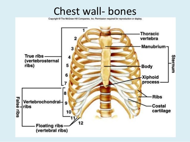
True, false and floating ribs are denoted. Surface anatomy of anterior chest wall. Pathology of the heart, mediastinum, lungs and pleura. It discusses the specific anatomy of the ribs and costal cartilages, along with the sternum. The anatomical structure of the 24 ribs in the human body is complex because of the irregular shape and different lengths of each rib.
Normal Female Anatomy Of The Chest Thoracic Cavity And Lungs Medical Art Works from cdn.shopify.com It originates at your clavicle, ribs, and sternum, and inserts into the upper portion of your humerus (upper arm. Among the major organs contained in the thoracic cavity are the heart and lungs. Spiral ct of thoracic inlet. ■ describe the anatomical relationships of various organs in the chest. It describes the theatre of events. The ribs are elastic arches of bone, which form a large part of the thoracic skeleton. It discusses the specific anatomy of the ribs and costal cartilages, along with the sternum. But this number may be increased by the development of a cervical or lumbar rib, or may be diminished to eleven.
Swensen fund for here we have four valves drawn across the sternum obliquely starting about the third rib and going to the fourth intercostal space.
As with all parts of the body, the anatomy and physiology of the chest wall are intimately intertwined. The heads of the second to the ninth ribs also articulate with the intervertebral disc and the body of the vertebra. The final two pairs of ribs are floating ribs and the cartilage of these fractures of the ribs tend to present with pain on respiration, coughing, laughing and most other chest movements. Insert contains images of a typical rib and the first rib. Ribs together form the rib cage, which as the name suggests, is a protective cage for the delicate thoracic organs such as lungs and heart. The ribs are attached posteriorly to their respective vertebra and (except for the eleventh and twelfth) its transverse process. Abnormalities of the rib cage include pectus excavatum (sunken chest) and pectus carinatum (pigeon chest). Right upper anatomy is to physiology as geography is to history: The chest anatomy includes the pectoralis major, pectoralis minor and the serratus anterior. It discusses the specific anatomy of the ribs and costal cartilages, along with the sternum. Human anatomy for muscle, reproductive, and skeleton. ■ describe the anatomical relationships of various organs in the chest. Identify the following structures on the lateral chest radiograph:
The ribs are elastic arches of bone, which form a large part of the thoracic skeleton anatomy of chest. Abnormalities of the rib cage include pectus excavatum (sunken chest) and pectus carinatum (pigeon chest).


0 Komentar